Content Overview
- Introduction
- Molecular and Pharmacological Background
- Mechanism of Action at the GABA-A Receptor
- Structure-Activity Relationships and Receptor Subtype Selectivity
- Laboratory and Preclinical Applications
- Radioligand and Imaging Studies
- Use in Behavioral and Electrophysiological Research
- Computational Modeling and Structural Analysis
- Methodological Considerations and Experimental Design
- Limitations and Confounding Factors
- Emerging Research Directions
- Summary
- Frequently Asked Questions (FAQ)
- References
Introduction
Flumazenil has become a cornerstone compound in neuropharmacological research. It’s best known as a selective benzodiazepine-site antagonist at the GABA-A receptor. Its combination of high affinity, selectivity, and reversibility has made it a go-to research tool for studying benzodiazepine receptor mechanisms. Although it first gained clinical recognition for reversing benzodiazepine-induced sedation, its scientific value reaches well beyond that use case. Researchers rely on it to study inhibitory neurotransmission, receptor density, and ligand binding kinetics across the central nervous system, without making any assumptions about therapeutic outcomes.
The GABAergic system, driven mainly by GABA-A receptors, plays a central role in keeping neuronal excitability in check. What makes flumazenil so useful is that it interacts with the benzodiazepine binding site without producing much intrinsic activity of its own. That gives researchers a clean way to dissect what’s happening at the receptor level. By competing with agonists and inverse agonists for the same site, it lets scientists probe the molecular structure and function of inhibitory signaling networks.
In the lab, flumazenil works as a probe, a comparator, and a standard reference ligand. It shows up in receptor binding assays, radiolabeling studies, electrophysiology experiments, and behavioral testing. Much of its versatility comes from having a well-characterized pharmacological profile, along with radiolabeled versions like [11C]-flumazenil and [3H]-flumazenil. This article walks through these areas in detail, covering research methods, the underlying mechanisms, and where the compound’s experimental limits lie.
Molecular and Pharmacological Background
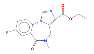
Flumazenil (chemically, ethyl 8-fluoro-5,6-dihydro-5-methyl-6-oxo-4H-imidazo[1,5-a]benzodiazepine-3-carboxylate) was developed in the late 1970s, during a period of intense research into benzodiazepine receptor antagonists. Its imidazobenzodiazepine structure closely resembles classical agonists like diazepam, but small differences in its electronic and steric properties change how it binds and how much intrinsic activity it produces.
The compound binds the benzodiazepine site with high nanomolar affinity but has almost no efficacy at the allosteric modulator site. In practice, that makes it a neutral competitive antagonist. Unlike inverse agonists such as beta-carbolines, flumazenil doesn’t change receptor-mediated chloride conductance on its own. It only blocks the effects of both agonists and inverse agonists when given alongside them.
These properties are exactly why flumazenil has been so useful for mapping benzodiazepine receptor occupancy, characterizing receptor subtype distribution, and studying how chronic ligand exposure changes receptor sensitivity. Because it can “unmask” baseline receptor activity without adding pharmacodynamic effects of its own, it’s particularly well suited to experimental pharmacology.
Mechanism of Action at the GABA-A Receptor
The GABA-A receptor is a heteropentameric chloride channel responsible for fast inhibitory signaling throughout the mammalian brain. It’s typically built from a mix of α, β, and γ (or δ) subunits, and that mix creates a wide range of receptor subtypes. Flumazenil binds selectively to receptors that contain a γ2 subunit paired with an α subunit of type α1, α2, α3, or α5.
At the benzodiazepine binding interface, located between the α and γ2 subunits, flumazenil goes head to head with allosteric modulators. Agonists like diazepam or midazolam boost GABA’s effect by increasing how often the channel opens. Inverse agonists do the opposite. Flumazenil, on the other hand, has little to no direct effect on channel gating. Instead, it simply blocks other ligands from binding in the first place.
This kind of competitive antagonism makes it possible to precisely measure receptor binding dynamics. When radiolabeled ligands get displaced by flumazenil in vitro, the resulting inhibition curves let researchers calculate binding affinity (Ki) and receptor density (Bmax). These numbers form the backbone of pharmacological modeling and receptor characterization work.
Structure-Activity Relationships and Receptor Subtype Selectivity
Structure-activity relationship (SAR) studies help explain why flumazenil is both highly selective and pharmacologically neutral. A fluorine group on the benzodiazepine ring boosts receptor affinity and makes the molecule easier to radiolabel. Meanwhile, the imidazole ring blocks the conformational shift that a full agonist would need to trigger.
Work using site-directed mutagenesis and cryo-electron microscopy has mapped out the specific amino acid residues that shape flumazenil binding. For example, structural studies of the synaptic GABA-A receptor bound to flumazenil show how substitutions within the α1 subunit, such as swapping histidine for arginine, can shift both binding strength and pharmacological neutrality. This kind of molecular detail has made flumazenil a benchmark ligand in structural neuropharmacology.
Subunit selectivity research suggests flumazenil is especially sensitive to receptors built from α1β2γ2 combinations, which dominate in cortical and limbic brain regions. That specificity gives researchers a useful proxy for tracking cortical inhibitory activity across development and disease states, without making any clinical diagnostic claims.
Laboratory and Preclinical Applications
Flumazenil shows up across several areas of preclinical and basic neuroscience research:
Receptor binding assays. By displacing radiolabeled benzodiazepine agonists, flumazenil helps define the size and affinity of a given receptor population.
Autoradiographic mapping. When radiolabeled, flumazenil can localize benzodiazepine binding sites across brain slices, revealing how inhibitory receptor density varies from one region to another.
Functional antagonism studies. Researchers use flumazenil to confirm that a behavioral or electrophysiological effect actually comes from benzodiazepine site activation, rather than some other form of GABA receptor modulation.
Comparative pharmacology. It acts as a neutral reference point against which agonists, partial agonists, and inverse agonists can all be tested.
PET and SPECT imaging. Radiolabeled flumazenil analogs make it possible to map receptors in vivo, in both animals and humans, for research purposes.
Studies using these methods have deepened our understanding of how inhibitory receptors are regulated during stress, learning, and other forms of neural adaptation. In particular, they’ve helped clarify how chronic benzodiazepine exposure changes receptor availability and desensitization, adding to the broader picture of synaptic plasticity.
Radioligand and Imaging Studies
Flumazenil’s usefulness in imaging research comes down to its radiochemical flexibility. The most widely used tracer, [11C]-flumazenil, is made by incorporating radioactive carbon into the molecule’s ethyl ester group, which gives it ideal properties as a positron emitter for PET scans.
Key research applications:
- In vivo receptor density mapping, which quantifies how much benzodiazepine binding site is available across cortical and subcortical brain regions.
- Pharmacokinetic modeling, used to work out rate constants for how quickly a ligand binds (kon) and unbinds (koff), which in turn informs receptor turnover.
- Pharmacological perturbation studies, which track receptor occupancy after giving a benzodiazepine-site ligand.
- Comparative species research, which allows side-by-side comparisons of GABAergic receptor distribution across rodents, primates, and humans.
Radiolabeled flumazenil’s moderate lipophilicity means it gets into the brain quickly and binds reversibly, which makes it well suited to dynamic scanning. For in vitro work, tritiated ([3H]) flumazenil is effective for detailed autoradiography and saturation binding studies.
That said, quantifying nonspecific binding and correcting for noise in reference-region input functions remains a real challenge in flumazenil PET imaging, which is why robust kinetic modeling protocols matter so much. Even with these challenges, flumazenil remains one of the most well-validated ligands available for GABAergic receptor imaging.
Use in Behavioral and Electrophysiological Research
In behavioral neuroscience, flumazenil’s neutral antagonist profile allows researchers to isolate exactly how much of an effect comes from the benzodiazepine receptor. Given systemically or directly into the brain in animal models, it can block benzodiazepine modulation without touching baseline GABAergic tone.
In electrophysiology experiments, flumazenil reverses the boost that benzodiazepine agonists give to inhibitory postsynaptic currents (IPSCs). That makes it possible to precisely measure benzodiazepine efficacy and how much receptor reserve is available.
In behavioral testing, flumazenil helps separate out anxiety-related effects that are specifically tied to the benzodiazepine site. For example, it can block an agonist’s tendency to increase open-arm exploration in the elevated plus-maze test, or counteract sedative effects in locomotor tests, all without producing much of an effect by itself.
Because of this, flumazenil acts as a critical control for identifying receptor-specific behavioral effects, and it’s given researchers real insight into how inhibitory modulation shapes cognition and emotion.
Computational Modeling and Structural Analysis
Recent computational work has added a lot to our understanding of how flumazenil interacts with the receptor at an atomic level. Molecular docking simulations and molecular dynamics analyses show how flumazenil holds the receptor in a neutral configuration, occupying the allosteric pocket without distorting it.
Homology models built from cryo-EM templates reveal that flumazenil sits differently depending on which α-subunit isoform is present. Researchers believe its weak hydrogen bonds with residues like α1H101 and γ2F77 explain why it binds reversibly and with high affinity, but without producing agonist-like effects.
Computational models are also being used to design new analogs, predicting derivatives with different lipophilicity, metabolic stability, or subtype preference. This work aims to develop better ligands for receptor imaging and neurochemical quantification, expanding the toolkit built around flumazenil.
Methodological Considerations and Experimental Design
Anyone using flumazenil in an experimental setup needs to account for a few key factors:
Dosage and concentration. In vitro work typically uses concentrations between 1 and 1000 nM for precise binding assays. In vivo dosing has to account for the drug’s fast hepatic metabolism and short plasma half-life.
Solvent compatibility. Because flumazenil doesn’t dissolve well in water, stock solutions usually need a DMSO or ethanol vehicle. Proper vehicle controls are essential to rule out solvent-related effects.
Receptor subtype diversity. Researchers should confirm receptor subunit composition when interpreting binding data, since α1-containing receptors are far more sensitive to flumazenil than α4- or α6-containing ones that lack a benzodiazepine site.
Time-course monitoring. Flumazenil’s fast onset and offset kinetics call for high temporal resolution in dynamic recordings.
Comparative ligand controls. Including diazepam, zolpidem, or beta-carbolines as controls helps confirm the validity of antagonism and reversibility testing.
Following these methodological principles helps keep results reproducible and ensures the data actually reflect what’s happening at the receptor level, rather than picking up systemic or pharmacokinetic noise.
Limitations and Confounding Factors
Despite its value, flumazenil comes with some real limitations:
Pharmacokinetic constraints. Fast metabolism and a short half-life make it hard to use in prolonged in vivo studies.
Intrinsic activity variability. Some expression systems have shown minor partial inverse agonist activity, possibly due to differences in receptor composition.
Off-target binding. At very high concentrations, flumazenil may weakly bind non-benzodiazepine GABA-A-like sites, which can complicate interpretation.
Species-specific differences. Rodent, primate, and human receptors don’t all bind flumazenil with the same affinity, so translating findings across species requires careful normalization.
Imaging quantification challenges. Nonspecific uptake and metabolite buildup complicate PET kinetics, often requiring arterial sampling or compartment modeling to sort out.
Keeping these caveats in mind is what allows flumazenil to stay a gold-standard ligand, while avoiding overinterpretation of what can be fairly complex receptor dynamics.
Emerging Research Directions
Advances in molecular technology keep expanding what flumazenil can do for research:
Cryo-EM and structural biology. High-resolution receptor structures are revealing how flumazenil’s presence changes local side-chain conformations, which gives researchers templates for designing new ligands.
Genetically encoded receptor tagging. Combining flumazenil binding assays with fluorescent or genetically encoded GABA receptor sensors opens the door to hybrid imaging approaches.
Radiochemistry innovations. New isotope-labeled analogs, such as [18F]-flumazenil variants, extend imaging half-life and improve resolution for preclinical PET studies.
Network-level analysis. Pairing flumazenil imaging with neural connectivity mapping helps link local receptor density to systems-level inhibitory control.
Machine learning integration. Predictive models of receptor occupancy and ligand-receptor energetics are being developed to forecast how flumazenil binds across different receptor subtypes and conditions.
Taken together, these developments point to flumazenil’s continuing role as a central tool in neuropsychopharmacological methodology, one that bridges molecular discovery and systems-level neuroscience.
Summary
Flumazenil is a foundational compound in neuropharmacology: an antagonist with no built-in therapeutic assumptions, but plenty of research value. Through its selective, competitive interaction with benzodiazepine binding sites on GABA-A receptors, it gives researchers a precise way to study inhibitory receptor architecture, function, and modulation.
Its reach extends from autoradiographic mapping to in vivo imaging, from molecular docking to behavioral testing. Despite some limitations tied to pharmacokinetics and receptor variability, flumazenil continues to set the methodological standard for probing GABAergic mechanisms.
By combining chemical neutrality with receptor specificity, flumazenil bridges molecular neuroscience and applied research, offering insight that’s fundamental to understanding synaptic inhibition and neurochemical balance.
Frequently Asked Questions (FAQ)
What is flumazenil used for in research? In laboratories, flumazenil works as a benzodiazepine-site antagonist used to study receptor binding, synaptic inhibition, and GABAergic modulation across different experimental systems.
How does flumazenil interact with GABA-A receptors? Flumazenil binds competitively at the benzodiazepine allosteric site. That blocks other benzodiazepine ligands from enhancing or suppressing the GABA response, without flumazenil itself directly activating the receptor.
What makes flumazenil suitable for imaging studies? When radiolabeled, flumazenil’s high-affinity, reversible binding allows researchers to visualize benzodiazepine receptor distribution and kinetics using PET or autoradiography.
Does flumazenil have any activity on its own? Under most conditions, it’s functionally neutral. Rare partial inverse effects have shown up in specific receptor configurations or at high concentrations, but this isn’t the norm.
What are the main experimental challenges with flumazenil? The biggest hurdles are its rapid metabolism, getting precise dosing right, avoiding nonspecific binding, and accounting for differences across receptor subtypes.
How does flumazenil compare to other antagonists? It’s one of the most selective and well-characterized antagonists at benzodiazepine sites, and it offers real advantages in specificity and radiolabeling compared with broader-acting GABAergic agents.
Can flumazenil derivatives provide new insights? Yes. Ongoing work on structural analogs and radiolabeled variants aims to improve receptor subtype targeting and imaging accuracy.
References
- Möhler, H., & Okada, T. (1977). Benzodiazepine receptor: Demonstration in the central nervous system. Science, 198(4319), 849-851. https://www.science.org/doi/10.1126/science.918669
- Hunkeler, W., Möhler, H., Pieri, L., Polc, P., Bonetti, E. P., Cumin, R., Schaffner, R., & Haefely, W. (1981). Selective antagonists of benzodiazepines. Nature, 290, 514-516. https://www.nature.com/articles/290514a0
- Sieghart, W., & Sperk, G. (2002). Subunit composition, distribution and function of GABA-A receptor subtypes. Current Topics in Medicinal Chemistry, 2, 795-816. https://www.eurekaselect.com/81522/article
- Olsen, R. W., & Sieghart, W. (2009). GABA-A receptors: Subtypes provide diversity of function and pharmacology. Neuropharmacology, 56(1), 141-148. https://pmc.ncbi.nlm.nih.gov/articles/PMC3525320/
- Lingford-Hughes, A., Hume, S. P., Feeney, A., Hirani, E., Osman, S., Cunningham, V. J., Pike, V. W., Brooks, D. J., & Nutt, D. J. (2002). Imaging the GABA-benzodiazepine receptor subtype containing the alpha5-subunit in vivo with PET. Journal of Cerebral Blood Flow & Metabolism, 22(7), 878-889. https://pubmed.ncbi.nlm.nih.gov/12142573/
- Selection of weighting factors for quantification of PET radioligand binding, including flumazenil, using simplified reference tissue models with noisy input functions. Journal of Cerebral Blood Flow & Metabolism. https://pmc.ncbi.nlm.nih.gov/articles/PMC3361066/
- Jacob, T. C. (2019). Neurobiology and therapeutic potential of α5-GABA type A receptors. Frontiers in Molecular Neuroscience, 12, 179. https://pmc.ncbi.nlm.nih.gov/articles/PMC6668551/
- Structure of a human synaptic GABA-A receptor, resolved by cryo-electron microscopy in complex with GABA and the benzodiazepine-site antagonist flumazenil. Nature. https://www.nature.com/articles/s41586-018-0255-3